Artlabeling Activities the Two Types of Cartilaginous Joints the Two Types of Cartilaginous Joints
Joints
Cartilaginous Joints
Learning Objectives
By the terminate of this section, y'all will be able to:
- Describe the structural features of cartilaginous joints
- Distinguish between a synchondrosis and symphysis
- Give an example of each type of cartilaginous joint
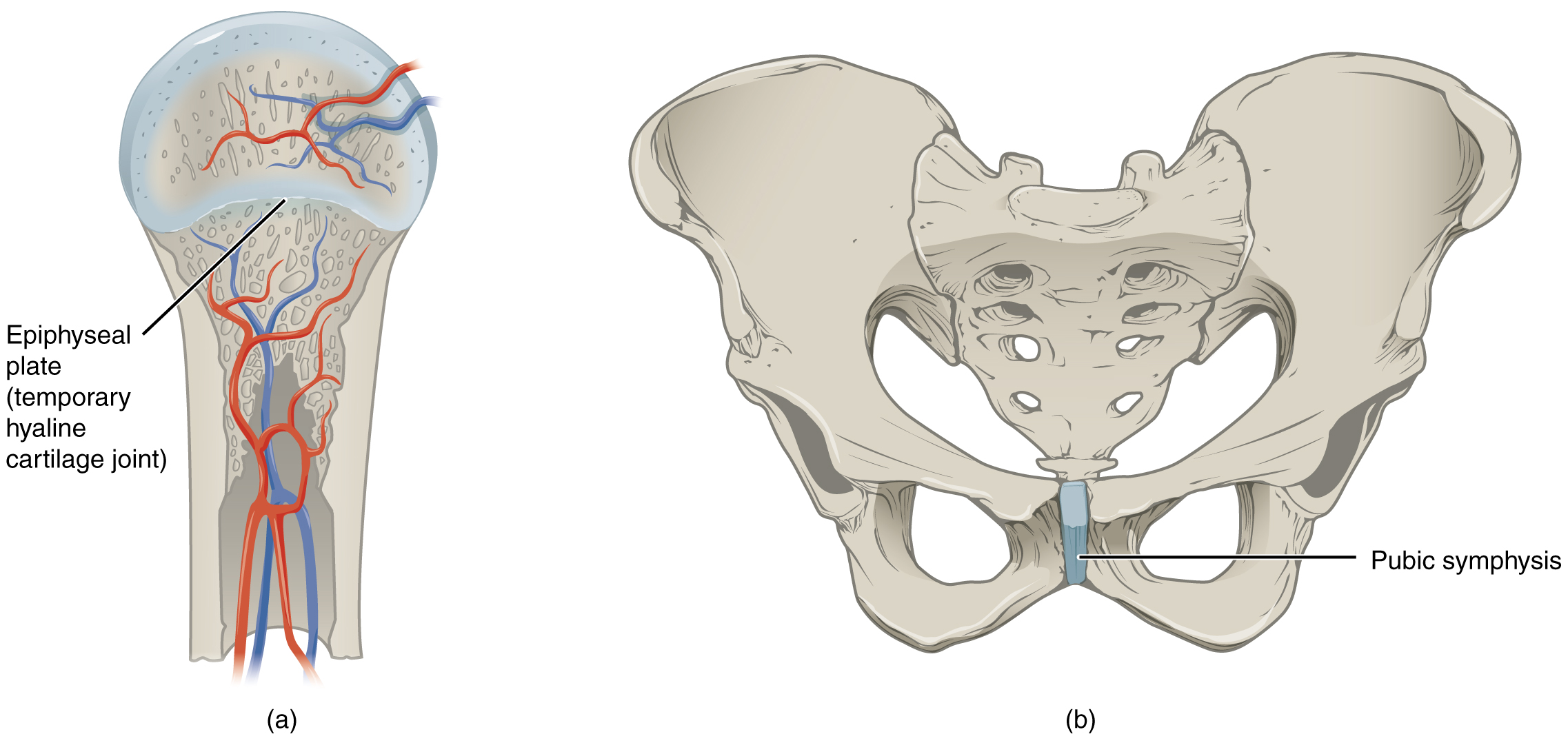
As the proper name indicates, at a cartilaginous articulation, the adjacent bones are united past cartilage, a tough but flexible blazon of connective tissue. These types of joints lack a articulation cavity and involve bones that are joined together by either hyaline cartilage or fibrocartilage ((Figure)). There are ii types of cartilaginous joints. A synchondrosis is a cartilaginous joint where the basic are joined by hyaline cartilage. Also classified equally a synchondrosis are places where bone is united to a cartilage structure, such every bit between the anterior end of a rib and the costal cartilage of the thoracic cage. The 2d type of cartilaginous joint is a symphysis, where the bones are joined past fibrocartilage.
Cartiliginous Joints
At cartilaginous joints, bones are united past hyaline cartilage to grade a synchondrosis or past fibrocartilage to form a symphysis. (a) The hyaline cartilage of the epiphyseal plate (growth plate) forms a synchondrosis that unites the shaft (diaphysis) and end (epiphysis) of a long os and allows the os to grow in length. (b) The pubic portions of the correct and left hip basic of the pelvis are joined together past fibrocartilage, forming the pubic symphysis.
Synchondrosis
A synchondrosis ("joined by cartilage") is a cartilaginous joint where bones are joined together by hyaline cartilage, or where bone is united to hyaline cartilage. A synchondrosis may be temporary or permanent. A temporary synchondrosis is the epiphyseal plate (growth plate) of a growing long os. The epiphyseal plate is the region of growing hyaline cartilage that unites the diaphysis (shaft) of the bone to the epiphysis (terminate of the bone). Bone lengthening involves growth of the epiphyseal plate cartilage and its replacement by bone, which adds to the diaphysis. For many years during childhood growth, the rates of cartilage growth and bone formation are equal and thus the epiphyseal plate does not change in overall thickness every bit the os lengthens. During the late teens and early on 20s, growth of the cartilage slows and eventually stops. The epiphyseal plate is then completely replaced past os, and the diaphysis and epiphysis portions of the bone fuse together to class a single adult bone. This fusion of the diaphysis and epiphysis is a synostosis. One time this occurs, bone lengthening ceases. For this reason, the epiphyseal plate is considered to be a temporary synchondrosis. Because cartilage is softer than bone tissue, injury to a growing long bone tin can damage the epiphyseal plate cartilage, thus stopping os growth and preventing additional bone lengthening.
Growing layers of cartilage also form synchondroses that bring together together the ilium, ischium, and pubic portions of the hip bone during babyhood and adolescence. When body growth stops, the cartilage disappears and is replaced by os, forming synostoses and fusing the bony components together into the unmarried hip bone of the adult. Similarly, synostoses unite the sacral vertebrae that fuse together to form the adult sacrum.
Visit this website to view a radiograph (X-ray paradigm) of a child's manus and wrist. The growing bones of child accept an epiphyseal plate that forms a synchondrosis betwixt the shaft and end of a long bone. Being less dense than bone, the area of epiphyseal cartilage is seen on this radiograph as the dark epiphyseal gaps located well-nigh the ends of the long basic, including the radius, ulna, metacarpal, and phalanx bones. Which of the bones in this prototype practise not show an epiphyseal plate (epiphyseal gap)?
Examples of permanent synchondroses are found in the thoracic cage. One case is the first sternocostal articulation, where the commencement rib is anchored to the manubrium by its costal cartilage. (The articulations of the remaining costal cartilages to the sternum are all synovial joints.) Boosted synchondroses are formed where the anterior end of the other eleven ribs is joined to its costal cartilage. Unlike the temporary synchondroses of the epiphyseal plate, these permanent synchondroses retain their hyaline cartilage and thus exercise not ossify with age. Due to the lack of move between the bone and cartilage, both temporary and permanent synchondroses are functionally classified as a synarthrosis.
Symphysis
A cartilaginous joint where the bones are joined by fibrocartilage is chosen a symphysis ("growing together"). Fibrocartilage is very stiff considering it contains numerous bundles of thick collagen fibers, thus giving it a much greater power to resist pulling and bending forces when compared with hyaline cartilage. This gives symphyses the power to strongly unite the next bones, but can nonetheless let for limited move to occur. Thus, a symphysis is functionally classified as an amphiarthrosis.
The gap separating the bones at a symphysis may be narrow or wide. Examples in which the gap between the bones is narrow include the pubic symphysis and the manubriosternal joint. At the pubic symphysis, the pubic portions of the right and left hip bones of the pelvis are joined together by fibrocartilage across a narrow gap. Similarly, at the manubriosternal joint, fibrocartilage unites the manubrium and body portions of the sternum.
The intervertebral symphysis is a wide symphysis located between the bodies of adjacent vertebrae of the vertebral column. Here a thick pad of fibrocartilage called an intervertebral disc strongly unites the next vertebrae past filling the gap between them. The width of the intervertebral symphysis is important considering information technology allows for small movements between the adjacent vertebrae. In addition, the thick intervertebral disc provides cushioning between the vertebrae, which is of import when carrying heavy objects or during high-bear upon activities such every bit running or jumping.
Chapter Review
There are two types of cartilaginous joints. A synchondrosis is formed when the adjacent bones are united by hyaline cartilage. A temporary synchondrosis is formed by the epiphyseal plate of a growing long bone, which is lost when the epiphyseal plate ossifies as the bone reaches maturity. The synchondrosis is thus replaced by a synostosis. Permanent synchondroses that do not congeal are found at the first sternocostal joint and between the inductive ends of the bony ribs and the junction with their costal cartilage. A symphysis is where the bones are joined past fibrocartilage and the gap betwixt the bones may be narrow or wide. A narrow symphysis is establish at the manubriosternal joint and at the pubic symphysis. A wide symphysis is the intervertebral symphysis in which the bodies of adjacent vertebrae are united by an intervertebral disc.
Interactive Link Questions
Go to this website to view a radiograph (X-ray image) of a child'due south hand and wrist. The growing bones of kid take an epiphyseal plate that forms a synchondrosis between the shaft and finish of a long bone. Being less dense than bone, the area of epiphyseal cartilage is seen on this radiograph equally the dark epiphyseal gaps located near the ends of the long bones, including the radius, ulna, metacarpal, and phalanx bones. Which of the basic in this image do not show an epiphyseal plate (epiphyseal gap)?
Although they are still growing, the carpal bones of the wrist area do non testify an epiphyseal plate. Instead of elongating, these basic abound in diameter past adding new bone to their surfaces.
Review Questions
A cartilaginous joint ________.
- has a joint crenel
- is called a symphysis when the bones are united by fibrocartilage
- anchors the teeth to the jaws
- is formed by a wide sail of gristly connective tissue
A synchondrosis is ________.
- found at the pubic symphysis
- where bones are connected together with fibrocartilage
- a type of fibrous joint
- plant at the start sternocostal joint of the thoracic cage
Which of the following are joined by a symphysis?
- adjacent vertebrae
- the kickoff rib and the sternum
- the terminate and shaft of a long bone
- the radius and ulna basic
The epiphyseal plate of a growing long bone in a child is classified every bit a ________.
- synchondrosis
- synostosis
- symphysis
- syndesmosis
Critical Thinking Questions
Draw the two types of cartilaginous joints and give examples of each.
Cartilaginous joints are where the adjacent bones are joined past cartilage. At a synchondrosis, the bones are united by hyaline cartilage. The epiphyseal plate of growing long bones and the first sternocostal articulation that unites the offset rib to the sternum are examples of synchondroses. At a symphysis, the bones are joined by fibrocartilage, which is stiff and flexible. Symphysis joints include the intervertebral symphysis betwixt adjacent vertebrae and the pubic symphysis that joins the pubic portions of the right and left hip bones.
Both functional and structural classifications can be used to depict an individual joint. Define the start sternocostal articulation and the pubic symphysis using both functional and structural characteristics.
The first sternocostal joint is a synchondrosis type of cartilaginous joint in which hyaline cartilage unites the starting time rib to the manubrium of the sternum. This forms an immobile (synarthrosis) blazon of joint. The pubic symphysis is a slightly mobile (amphiarthrosis) cartilaginous articulation, where the pubic portions of the right and left hip bones are united past fibrocartilage, thus forming a symphysis.
Glossary
- symphysis
- type of cartilaginous joint where the bones are joined past fibrocartilage
- synchondrosis
- blazon of cartilaginous articulation where the bones are joined by hyaline cartilage
hastingsfuser2001.blogspot.com
Source: https://opentextbc.ca/anatomyandphysiologyopenstax/chapter/cartilaginous-joints/
0 Response to "Artlabeling Activities the Two Types of Cartilaginous Joints the Two Types of Cartilaginous Joints"
Post a Comment